Author Archives: Ashley Mota

Reflection on Myself as a Writer

At the beginning of the semester, the class was asked to write a reflection answering four questions: What are your strengths as a writer? What are your weaknesses as a writer? What are your reading and editing strategies? What is your experience as a writer? What do you hope to take from this semester? The following was my reflection on myself as a writer. Every thing that I mentioned in this reflection has in one way or another appeared in my other works in the rest of my portfolio.

I have always felt that writing is not my strong suit. I often struggle to even begin writing things like essays, because I don’t know where to start or how to articulate my words – honestly I sometimes even lack the motivation. However, once I find the right words to start with, I tend to go over the word count and instead face the weakness of not being concise. Some strengths I hold are that I enjoy using transitional words, phrases, or even sentences, because I like to make my work flow. Additionally, I like to explore with creative styles of writing like letters or made-up stories, full of imagery.

When it comes to reading and editing strategies, I need to work on them more, because after I write an essay or a writing piece of some sort, I do not usually go back and edit it. I read it over to check for spelling and grammar, but never to see if I wrote too much or too little, not do I edit its content. My experience as a writer has been purely academic. I have written research papers and essays for school, lab reports for biology, chemistry, and physics, and poems for creative writing assignments. This semester, I hope to take a lot away from this course. My goals are to improve upon my editing skills- to help both myself and my peers- to strengthen my ability to write thesis statements that eloquently reflect my stance and main ideas, and also to learn more about the different types of writing styles available. Overall, I am aiming to continue growing and improving my writing skills by learning more about my writing style- both my strengths and my weaknesses.

Literature Review Draft

The following document is the draft for my literature review. In order to organize my thoughts, I used a bullet point outline format. With the ideas for my headers nearly set in stone, it was easy for me to go through the articles and pinpoint specific areas from each one that fit into the specific heading. The abstract was left empty because that was the last paragraph that was written for this piece.

Misdiagnosis of Epilepsy: A Literature Review

ABSTRACT

INTRODUCTION

Epilepsy is a serious condition thatwhich has different symptoms, among which seizure is the most common. The disturbance in brain activity that is caused by the symptoms of epilepsy can be harmful to the body and brain. Therefore, once an epileptic diagnosis is made, actions for treatment should be taken right away. Epilepsy can be treated with differentTreatment for epilepsy can be different anti-epilepsy medication. While epilepsy is a very serious condition, there are accounts of common misdiagnosis of epilepsy. Misdiagnosis of epilepsy can occur in two waysbe seen in two forms: one is diagnosing a condition as epilepsy, when it is not, or diagnosing a different condition as nother condition when it is epilepsy. In this literature review, multiplea different number of studies will be analyzed and compared to target which factors contribute to the common misdiagnosis of epilepsy. The aim is to pinpoint those factors and identify a possible solution so that the rate of misdiagnosis can be reduced. This topic is of great importance because a misdiagnosis of epilepsy can have many detrimentalundiscovered implications for the patient receiving the wrong diagnosis. When thinking of the importance of treatment for epilepsy, a possible implication of misdiagnosis is that a person will not receive the proper treatment for their conditionthe treatment they possess- whether it be epilepsy or another condition. Additionally, a person beingcan be treated for epilepsy will suffer unwanted side effects from the medication which can possibly be detrimental to their healthan illness they do not have and experience unknown side effects. With the analysis of the main factors leading to the misdiagnosis of epilepsy, the implications can be known, and a possible solution can be established.

ANALYSIS

Common Non-Epileptic Events Confused with Epilepsy

·     Syncope is the most common non-epileptic event that is mistaken with epilepsy (Hindley 2006; Fattouch et al 2007).

Lack of Knowledge about Different Symptoms *These two might work hand in hand, because it deals with differing symptoms that can be confused with each other*

Misinterpretation of Clinical Data: EEGs, MRI, and Patient History  

·     The article states that getting a good reading on an EEG is very difficult in clinical setting that are not specialized in epilepsy. (Uldall et al 2006)

·     21 out of the 30 SMS patients showed abnormal EEGs and 11 out of 30 showed some alteration in their MRI findings. In the US group, 9 out of 27 had EEG abnormality and only 1 had an MRI alteration. (Fattouch 2007)

Doctors’ Certainty of Diagnosis:

·     The certainty of a doctor on the diagnosis they are giving can deter from the right diagnosis being given (*alternate sentence: … can deter from the proper steps being taken to reach confirmation of an intended diagnosis).

o  Some doctors underestimate symptoms because they don’t want to give a harsh diagnosis such as epilepsy for symptoms they are unclear of, to then have to retract it. So they prefer to label it as not being epilepsy

CONCLUSION

Understood Implications?

*Maybe this should be my conclusion?* Possible Solution: Referrals to epilepsy centers before making a definite diagnosis

·     that centers that do not specialize in epilepsy will have a harder time in properly diagnosing epilepsy and syncope because of the misinterpretation of clinical, EEG, and neuroimaging results. (Fattouch 2007)

·     The article states that getting a good reading on an EEG is very difficult in clinical setting that are not specialized in epilepsy. (Uldall et al 2006)

o  Since misinterpretation of EEGs and other clinical tests might have occurred or are very difficult in areas not specialized for epilepsy, then it can be inferred that a possible solution to the misdiagnosis of epilepsy is the referral to epilepsy centers before an epilepsy diagnosis is made.

·     “if the cause of a ‘funny turn’ is unclear, it is better to err on the side of ‘not epilepsy’ and subsequently correct that decision than to give the label of ‘epilepsy’ and try to retract it”.( O’Donohoe 1994 cited by Hindley 2006)

References:

Fattouch J., Di Bonaventura C., Strano S., Vanacore N., Manfredi M., Prencipe M., Giallonardo A.T. 2007. Over-interpretation of electroclinical and neuroimaging findings in syncope misdiagnosed as epileptic seizures. Epileptic Discord. 9(2)(170-173) [Internet][Accessed May 8 2019]

Available from: https://pdfs.semanticscholar.org/d2f5/2e5e508d5cb5fbae52f1d0256a4759220345.pdf

Hindley D., Ali A., Robson C. 2006. Diagnoses made in a secondary care “fits, faints, and funny turns” clinic. ADC. 91(3)(214-218). [Internet] [Accessed April 15 2019]

Available from: https://adc-bmj-com.clinical-proxy.libr.ccny.cuny.edu/content/91/3/214

Murthy JM. 1999. Factors of error involved in the diagnosis of juvenile myoclonic epilepsy: a study from South India. Neurology India. 47(3)(210-213). [ Internet] [Accessed April 13 2019]

Available from: http://www.neurologyindia.com/article.asp?issn=0028-3886;year=1999;volume=47;issue=3;spage=210;epage=3;aulast=murthy

Uldall P., Alving J., Hansen L K., Kibaek M., Buchholt J. 2006. The misdiagnosis of epilepsy in children admitted to a tertiary epilepsy centre with paroxysmal events. ADC. 91(3)(219-221). [Internet] [Accessed April 10 2019]

Available from: https://adc-bmj-com.clinical-proxy.libr.ccny.cuny.edu/content/91/3/219

Literature Review Assignment

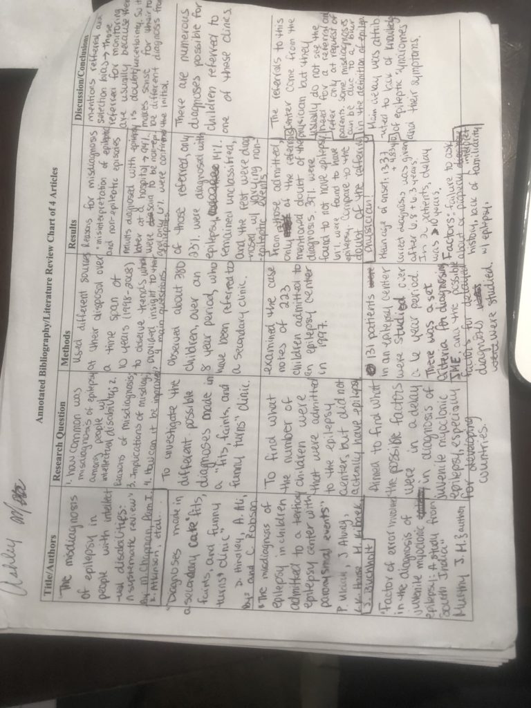
This assignment was a Literature Review. I chose the topic of “misdiagnosis of epilepsy” after I was it mentioned in a pediatric journal. In this assignment I reviewed 4 different articles to explore the main factors which led to the common misdiagnosis of epilepsy. Once I found the main components listed, in nearly all of the articles, I chose those factors as the subheadings for the Analysis portion of the paper. Analysis of those factors allowed for an inference to what the solution to the problem of misdiagnosing epilepsy, which was the basis of the conclusion.

Factors Resulting in the Misdiagnosis of Epilepsy: A Literature Review

ABSTRACT

            The condition of epilepsy is one that has many different symptoms and is commonly misdiagnosed. Epilepsy misdiagnosis can be seen through labeling a non-epileptic condition as epilepsy or an epileptic condition being labeled as being non-epileptic. The major factors leading to misdiagnosis are 1) confusion between epileptic symptoms and non-epileptic episodes, 2) misinterpretation of the results from different clinical tests such as EEGs and MRIs, and 3) the lack of referral to epileptic centers based on the certainty of doctors in their initial diagnoses. The misdiagnosis of epilepsy has a major implication for the treatment which patients receive- in which they might be getting inappropriate treatment for a condition they do not possess. A possible solution to lessen the extent to which epilepsy is misdiagnosed can be to always refer epileptic-like cases to a center which specializes in epilepsy, so that a diagnosis is more accurate.  

INTRODUCTION

 Epilepsy is a serious condition which has different symptoms, among which seizure is the most common. The disturbance in brain activity that is caused by the symptoms of epilepsy can be harmful to the body and brain. Therefore, once an epileptic diagnosis is made, actions for treatment should be taken right away. Treatment for epilepsy can be different anti-epilepsy medication. While epilepsy is a very serious condition, there are accounts of common misdiagnosis of epilepsy. Misdiagnosis of epilepsy can be seen in two forms: one is diagnosing a condition as epilepsy, when it is not, or diagnosing another condition when it is epilepsy. In this literature review, a different number of studies will be analyzed and compared to target which factors contribute to the common misdiagnosis of epilepsy. The aim is to pinpoint those factors and identify a possible solution so that the rate of misdiagnosis can be reduced. This topic is of great importance because a misdiagnosis of epilepsy can have many undiscovered implications for the patient receiving the wrong diagnosis. When thinking of the importance of treatment for epilepsy, a possible implication of misdiagnosis is that a person will not receive the proper treatment for the treatment they possess- whether it be epilepsy or another condition. Additionally, a person can be treated for an illness they do not have and experience unknown side effects. With the analysis of the main factors leading to the misdiagnosis of epilepsy, the implications can be known, and a possible solution can be established.

ANALYSIS

Common Non-Epileptic Events and Symptoms Confused with Epilepsy

There are many different diagnosis possibilities that can be assigned to symptoms common to epilepsy, among which some were syncope, migraines, daydreaming, night terrors, psychological episodic spells, tics, staring, dystonia, and disturbances during sleep (Hindley et al 2006; Uldall et al 2006). Syncope is the most common non-epileptic event that is mistaken with epilepsy (Hindley et al 2006; Fattouch et al 2007). Two of the most notable differences between epileptic events and non-epileptic events were that non-epileptic events were situational, meaning that they were triggered by a specific event or activity, and that non-epileptic episodes could be interrupted (Hindley et al 2006).This shows that even if symptoms seem to be the same for epileptic and non-epileptic episodes, there are other factors that could come into play which could aid in differentiating the two. Fattouch (2007) supported this idea with the use of a questionnaire and scoring system that differentiated between epileptic seizures and syncope. The questionnaire asked different questions and each question had a specific scoring where it added points or subtracted points. Some of the questions were if there was unresponsiveness during the spell, if there was sweating before the spell, and if the spell was associated with prolonged sitting or standing. The question regarding unresponsiveness during a spell gets a point, going towards the characteristics of an epileptic seizure, and sweating before a spell or the spell being caused by prolonged sitting or standing loses two points, going away from epileptic seizure and moving towards a syncope diagnosis. These three characteristics depict examples of how to distinguish between non-epileptic and epileptic spells using two of the differentiating factors expressed by Hindley (2006).

These cases dealt with an epileptic diagnosis being given to non-epileptic events. However, the reverse can happen as well. The lack of knowledge regarding the symptoms of epilepsy can lead to an improper diagnosis of an actual epileptic episode. In other words, symptoms might be labeled as being non-epileptic when in reality the patient possesses epilepsy. Murthy (1999) conducted a study for the factors leading to delay in diagnosis of Juvenile Myoclonic Epilepsy (JME) in South India with 131 patients. The symptoms for JME are explicitly known; however, the time span in which they appear can vary between each other. For example, generalized tonic clonic seizures (GTCS) are often the first symptoms to appear but the circadian relations to awakening from sleep might not be fully manifested right away. Additionally, the appearance of absence seizures can precede GTCS and myoclonic jerks by 4 to 5 years. If a doctor is unaware of the time span and characteristics of the symptoms of JME, they might just give a diagnosis of absence epilepsy and not even consider JME as a potential future problem. If the contrasts between non-epileptic episodes and specific epileptic seizures are not very well known, there can be an effect on the diagnosis given to a patient.

Misinterpretation of Data: EEGs, MRI, Etc.

When in centers that do not specialize in epilepsy, it is hard to accurately analyze the data collected through different tests in order to give the proper diagnosis (Britton 2004 cited by Fattouch et al 2007; Uldall et al 2006).  The study conducted by Fattouch et al (2007) had a cohort of 62 subjects, 57 of which were found to have a definite diagnosis of syncope. They were divided up into two groups to further analyze their data accordingly. 30 of the patients had received a “definite epileptic” diagnosis; they made up the syncopes misdiagnosed as epileptic seizures (SMS) group. 27 of the patients were only suspected of having epilepsy; these individuals were labeled as unrecognized syncopes (US). 70% of the SMS patients showed abnormal EEGs and 37% showed some alteration in their MRI findings. In the US group, 33% had EEG abnormality and only 1 had an MRI alteration. These statistics showed that there were abnormal results collected from the MRI and EEGs, however, all of the subjects had a syncope diagnosis, despite those abnormalities.  

Similarly, in Hindley’s (2006) study, of 380 subjects, there were differing abnormalities found. Abnormalities were reported in 84% of the subject count. Of this group, 85% of them were abnormal at the first testing, but for 15% a repeat EEG, sleep EEG, or prolonged EEG was required. This showed that due to uncertainty, a second testing might be needed for established accuracy.  53% of the entire subject population had computed tomography (CT) scans, out of which 23% showed some type of abnormality. 52% had magnetic resonance imaging (MRI) scans, and 41% of this group showed abnormality. The large presence of abnormalities could have misled an epileptic diagnosis. However, only 23% of the subject population was actually confirmed to have an epilepsy diagnosis.  

This shows that some doctors might be tricked by those abnormalities thinking that they point directly to an epileptic characteristic of EEGs and MRIs. However, a proper analysis is required to be able to distinguish between abnormal syncope, or other non-epileptic events, results and definite epileptic results.

Doctors’ Certainty of Diagnosis:

The certainty of a doctor on the diagnosis they are giving can deter from the proper steps being taken to reach confirmation of an intended diagnosis. Some doctors underestimate symptoms because they don’t want to give a harsh diagnosis such as epilepsy for symptoms, which they are unclear of, to then have to retract it. So, they prefer to label it as not being epilepsy, and then correct it after the fact. (O’Donohoe 1994 cited by Hindley 2006) In Uldall’s (2006) et al study, 223 children were observed, 17% of them had referrals where their doctor had doubt about their epileptic diagnosis. From these uncertain referrals, 18% had confirmed epilepsy and 82% did not have confirmed epilepsy. These doctors, worked similar to what Hindley talked about, showing that it was better to have a non-epileptic or uncertain diagnosis to then have it corrected. In contrast to this idea, Uldall’s (2006) study also had 83% of their subject population with referrals where their doctors were “without a doubt” certain of their epileptic diagnosis. From these, 70% had a confirmed epileptic diagnosis and 30% did not actually have epilepsy. The referrals from these doctors came mostly at the request of parents. This shows that doctor’s could have such a strong feeling about their epileptic diagnosis, despite their equipment and setting being inferior to that of the epileptic centers, that 30% of patients would have been dismissed with the wrong diagnosis. This is the perfect depiction of when the certainty of a doctor could negatively impact the misdiagnosis of epilepsy.

CONCLUSION

            When dealing with the diagnosis of epilepsy, it is important to take into consideration the different symptoms and possible non-epileptic diagnoses, as well as accurately decipher the clinical data before a final diagnosis is made (Murthy 1999; Hindley et al 2006; Uldall et al 2006; Fattouch et al 2007). Additionally, doctors should never be too certain in their diagnosis, especially one that was made outside of an epileptic center. Since major factors established to affect misdiagnosis of epilepsy are often present in areas not specialized for epilepsy, it can be inferred that a proposed solution to the misdiagnosis of epilepsy is the referral of any episodes that even seemingly resemble epilepsy to epilepsy centers before an epilepsy diagnosis is made (Murthy 1999; Hindley et al 2006; Uldall et al 2006; Fattouch et al 2007). The major implications for the misdiagnosis of epilepsy can be a delay in appropriate treatment for the proper condition. The exact side effects of receiving treatment for the wrong condition was not explored by these researchers; therefore, that is an area for possible future directions in this topic.

References:

Fattouch J., Di Bonaventura C., Strano S., Vanacore N., Manfredi M., Prencipe M., Giallonardo A.T. 2007. Over-interpretation of electroclinical and neuroimaging findings in syncope misdiagnosed as epileptic seizures. Epileptic Discord. 9(2)(170-173) [Internet][Accessed May 8 2019]

Available from: https://pdfs.semanticscholar.org/d2f5/2e5e508d5cb5fbae52f1d0256a4759220345.pdf    

Hindley D., Ali A., Robson C. 2006. Diagnoses made in a secondary care “fits, faints, and funny turns” clinic. ADC. 91(3)(214-218). [Internet] [Accessed April 15 2019]

Available from: https://adc-bmj-com.clinical-proxy.libr.ccny.cuny.edu/content/91/3/214

Murthy JM. 1999. Factors of error involved in the diagnosis of juvenile myoclonic epilepsy: a study from South India. Neurology India. 47(3)(210-213). [ Internet] [Accessed April 13 2019]

Available from: http://www.neurologyindia.com/article.asp?issn=0028-3886;year=1999;volume=47;issue=3;spage=210;epage=3;aulast=murthy

Uldall P., Alving J., Hansen L K., Kibaek M., Buchholt J. 2006. The misdiagnosis of epilepsy in children admitted to a tertiary epilepsy centre with paroxysmal events. ADC. 91(3)(219-221). [Internet] [Accessed April 10 2019]

Available from: https://adc-bmj-com.clinical-proxy.libr.ccny.cuny.edu/content/91/3/219

Annotated Bibliography Chart of Articles

The following image is a chart with headings: title/authors, research question, methods, results, and discussion/conclusion. This chart was used to separate each article into the above headings. By separating them in the columns, it was easier to compare them to each other and see the differences and similarities in them. This chart was a very useful organization tool in constructing my annotated bibliography because I had already listed the main points of each section of the articles.

Annotated Bibliography Draft

The below work is the draft of an annotated bibliography which was then used to write a literature review. This draft was not a very effective form for the assignment, because it did not allow for sufficient feed back in all parts associated with writing an annotated bib. The draft only started to speak of the main points of each article, but I did not include any of the credibility evaluations nor the purpose of the article for the literature review. Therefore, the feedback I received was only on the ability to begin summarizing an article and not on its effective use nor the analysis of its credibility.

Misdiagnosis of Epilepsy: An Annotated Bibliography

Chapman M., Iddon P., Atkinson K., Brodie C., Mitchell D., Parvin G., and Willis S. 2011. The misdiagnosis of epilepsy in intellectual disabilities: a systematic review. Seizure. 20(2)(101-106) [Internet][Accessed April 10 2019]

Available from: https://www.sciencedirect.com/science/article/pii/S105913111000261X

This article addresses the concept of misdiagnosis involving epilepsy, especially in patients with intellectual disabilities, a population in which epilepsy can be very common. The questions this study aimed to answer were: how common the misdiagnosis of epilepsy in individuals with intellectual disabilities was, what were the reasons for the misdiagnosis, what were the implications of the misdiagnosis, and how can future diagnostics be improved.

Hindley D., Ali A., Robson C. 2006. Diagnoses made in a secondary care “fits, faints, and funny turns” clinic. ADC. 91(3)(214-218). [Internet] [Accessed April 15 2019]

Available from: https://adc-bmj-com.clinical-proxy.libr.ccny.cuny.edu/content/91/3/214

This article deals with children referred to a secondary clinic from different sources- pediatricians, psychiatrists, pediatric neurologists- with an epilepsy diagnosis. However, there were varying symptoms in the children which could have been from a range of different diagnosis. 23% were reinforced an epileptic diagnosis, 43% had a diagnosis of syncope, 36% had some other non-epileptic event as their diagnosis, and 14% were unclassified. This article presents different paroxysmal events present in infancy and childhood which are very common and can often be confused with epilepsy- leading to the commonality of misdiagnosing certain symptoms as being epileptic.

Murthy JM. 1999. Factors of error involved in the diagnosis of juvenile myoclonic epilepsy: a study from South India. Neurology India. 47(3)(210-213). [ Internet] [Accessed April 13 2019]

Available from:http://www.neurologyindia.com/article.asp?issn=0028-3886;year=1999;volume=47;issue=3;spage=210;epage=3;aulast=murthy

This article discusses the misdiagnosis of epilepsy in a developing country- South India- and has a focus on the time stamp of diagnosis more than the incorrect labeling of epilepsy (which differs from the other articles). Murthy aimed to find some possible reasons as to why Juevnile Myoclonic epilepsy (JME) is so often underdiagnosed or diagnosed very late after arise of first symptoms. The time stamp was found to be an average of 7 years, and as late as 10+ years in 20 patients.

Uldall P., Alving J., Hansen L K., Kibaek M., Buchholt J. 2006. The misdiagnosis of epilepsy in children admitted to a tertiary epilepsy centre with paroxysmal events. ADC. 91(3)(219-221). [Internet] [Accessed April 10 2019]

Available from: https://adc-bmj-com.clinical-proxy.libr.ccny.cuny.edu/content/91/3/219

In this article, there was an extensive review of case notes regarding 223 children admitted to an epilepsy center. About 86% of the children admitted were already on antiepileptic drugs, which could have had a detrimental effect on those who did not actually have epilepsy.  Out of those that came in with a possible epileptic diagnosis, 39% were found to not actually have epilepsy, and those who were supposedly “without any doubts” to be epileptic, 30% of them had their diagnosis disproven. About 47% of the children who were admitted as first timers with varying symptoms were quickly discharged as presenting with non-epileptic seizures.

Annotated Bibliography Original

This final copy of my annotated bibliography had a source which was not original research, but a systematic review, for that reason it is labeled the original. However, it is not the final copy which I received a grade for in class. This annotated bibliography was effective in summarizing the important parts of the article and listing what their functions would be in the literature review. The part I struggled with most for this assignment was the credibility of the article, I had to search for some outside sources to find the credentials of the authors. For most of the authors however I couldn’t find much information.

Misdiagnosis of Epilepsy: An Annotated Bibliography

Chapman M., Iddon P., Atkinson K., Brodie C., Mitchell D., Parvin G., and Willis S. 2011. The misdiagnosis of epilepsy in intellectual disabilities: a systematic review. Seizure. 20(2)(101-106) [Internet][Accessed April 10 2019] Available from: https://www.sciencedirect.com/science/article/pii/S105913111000261X

This article addresses the concept of misdiagnosis involving epilepsy, especially in patients with intellectual disabilities, a population in which epilepsy can be very common. The questions this study aimed to answer were: how common the misdiagnosis of epilepsy in individuals with intellectual disabilities was, what were the reasons for the misdiagnosis, what were the implications of the misdiagnosis, and how can future diagnostics be improved. One of the main points made in this article refers to the negative implications that can befall a patient once misdiagnosed with epilepsy. The implications stated were distress to both the patient and those taking care of them, any social stigmatization surrounding epileptic people, financial deprivation, and unnecessary treatment. 48% of patients with Rett Syndrome had seizures that were not associated with EEG epileptic seizures but were still receiving antiepileptic medication. On the flipside, patients in need of epileptic treatment might not receive it because they have not been properly diagnosed- 30% of women with Rett Syndrome were in need of antiepileptic medication but were not receiving it. A large part of misdiagnosis was said, in this article, to come from the inability of parents, caregivers, and/or health professionals to accurately interpret signs as being epileptic or non-epileptic events. The results showed that about 82% of parents incorrectly reported an epileptic episode in their daughters with Rett Syndrome. Whereas there were 13 individuals with Rett Syndrome who actually had epileptic symptoms based on their EEGs, but only 5 of those 13 parents were able to identify the episode as an epileptic one. Poor training on the part of health care professionals can often lead to a misdiagnosis of epilepsy. Along with poor training there might have been cases of poor communication and inconsistency in checking on the patient. It was stated that these factors leading to misdiagnosis can be most prominent when dealing with rare disability syndromes, because these symptoms aren’t often very known and can be mistaken for epileptic events. An EEG is one tool used for the diagnosis of epilepsy, however, in the conclusion it is stated that an EEG was not always used. When used it was not determined if the length of time designated for the EEG was enough to detect epileptic seizures. Another very strong point was made in the conclusion of this article, stating that when a patient is referred for monitoring, there is some type of doubt associated with their initial diagnosis and uncertainty with what the right diagnostic would be. Therefore, it makes sense for there to be an overestimation to then be referred for monitoring. This article will be useful for the literature review to present data and important points referring to what a misdiagnosis can cause in patients as well as some pressing factors which might lead to a misdiagnosis or overestimated diagnosis.

            An evaluation of the credibility of this article pointed towards it being a credible source due to the diversity of skills and knowledge of its authors. Also, the different databases were reviewed by at least two different authors to ensure there was no bias or misinterpretation occurring. Melanie J Chapman is a Faculty of Health, Psychology, and Social Care at Manchester Metropolitan University with a specialty in intellectual disabilities. This shows that she is very qualified to speak on the issue of misdiagnosis and treatment of those with intellectual disabilities.

Hindley D., Ali A., Robson C. 2006. Diagnoses made in a secondary care “fits, faints, and funny turns” clinic. ADC. 91(3)(214-218). [Internet] [Accessed April 15 2019]Available from: https://adc-bmj-com.clinical-proxy.libr.ccny.cuny.edu/content/91/3/214

This article deals with 380 children referred to a secondary clinic from different sources- pediatricians, psychiatrists, pediatric neurologists- with an epilepsy diagnosis over an eight-year period (1995-2003). The children referred to these clinics were either first timers, showing symptoms for the first time, or those that already had an epileptic diagnosis and needed further specification for management. The diagnoses were made based on family history, any previous clinical information, and the witnessed accounts of parents and family-members (these were preferably taped, which allowed for interpretation of the doctor instead of from the parents who had no idea what they were witnessing). The results showed that 23% (89 children) were newly diagnosed with epilepsy, 26% (100 children) were diagnosed with syncope, 36% (138 children) were diagnosed with other non-epileptic events, and 14% (53 children) were labeled as unclassified events. In those diagnosed with non-epileptic events, different paroxysmal events were present, among which identified were syncope, psychological episodes, daydreams, night terrors, migraines, benign paroxysmal vertigo, ritualistic movements, and exaggerated parental anxiety or fabricated illness. All of these are very common in infancy and childhood and can often be confused with epilepsy. For example, frequent daydreams can be referred to an epileptic specialist out of fear that a child might be experiencing something known as childhood absence epilepsy. The commonalities between the non-epileptic and epileptic symptoms can often cause the misdiagnosis of epilepsy by viewing any unusual mannerisms and behaviors. This article will be used in the literature review as a source of different non-epileptic diagnostics available that present similar to epileptic symptoms and contribute to the common misdiagnosis of epilepsy.

When establishing the credibility of this article, the time span across which it takes place was considered. The fact that the data was collected over a time span of eight years gives it an aspect of accuracy because it had a relatively large sample size and varying time stamps. An aspect which gives the experiment itself credibility is the fact that the same clinician saw all of the children used in this study. This adds a layer of credibility because there is no discrepancy or area for opposing views because it was all performed by one doctor. All three authors of this article are associated with Fairfield General Hospital and the University of Manchester. Dr. D Hindley is a consultant Pediatrician at Halliwell Children’s Center which contributes to their credibility to speak about children and their behaviors which could be confused with epileptic symptoms.

Murthy JM. 1999. Factors of error involved in the diagnosis of juvenile myoclonic epilepsy: a study from South India. Neurology India. 47(3)(210-213). [ Internet] [Accessed April 13 2019] Available from: http://www.neurologyindia.com/article.asp?issn=0028-3886;year=1999;volume=47;issue=3;spage=210;epage=3;aulast=murthy

This article discusses the misdiagnosis of epilepsy, specifically of Juvenile Myoclonic Epilpesy, in a developing country- South India. It has a focus on the time stamp of diagnosis rather than the incorrect labeling of epilepsy. Murthy aimed to find some possible reasons as to why Juvenile Myoclonic Epilepsy (JME) is so often underdiagnosed or diagnosed very late after rise of first symptoms. 131 patients were studied, 90% of those patients were immediately diagnosed at the time of registration at the clinic and 10% were diagnosed during a follow-up. Th article lists the median age of onset as 13.37 + 4.83 years old, and that it took 6.8 + 6.3 years from the onset of the disease for it to be diagnosed properly. In 20 of the patients (15%), it took more than 10 years for diagnosis. Murthy mentions that major factors of delay in diagnosis were the failure to ask about or properly interpret the history of the presence of myoclonic jerks and the lack of knowledge regarding the symptoms of this specific epileptic syndrome. Another factor is that patients might go to the clinic looking for treatment of a specific type of seizure which causes a delay in doctors properly diagnosing the presence of JME. For example, 9 patients had an initial diagnosis of absence epilepsy and the seizures they were seeking treatment for were absences, however their EEGs showed wave activity in conjunction with JME criteria. Additionally, there is the possibility to recognize one of the JME criteria symptoms but not another, making it harder to fully diagnose JME. For example, generalized tonic clonic seizures (GTCS) are often the first symptoms to appear but the circadian relations to awakening from sleep might be fully manifested right away, two symptoms which are complementary of JME. One important thing pointed out in this article is that some criteria of JME might make an appearance years in advance before another, such as the appearance of absence seizures can precede GTCS and myoclonic jerks by 4 to 5 years. Once the myoclonic jerks and GTCS are present is when the official diagnosis of JME can be established. Meaning that some patients might receive an preliminary childhood or juvenile absence epilepsy before it progresses to a JME diagnosis. This article will pose as record of factors which can both attribute to misdiagnosis as well as delay in diagnosis of epilepsy, specifically of JME. It can also serve as a basis for why constant follow-ups are important, especially when an initial epilepsy diagnosis is given.

Dr. J M Murthy is a Neurologist in hospital care in India. Her areas of specialty are neuro electrophysiology, movement disorders, neuro muscular disorders, tumors, spine and spinal disorders, migraines, strokes, and epilepsy. This attests to her ability to discuss matters concerning epilepsy diagnosis, EEG readings, and care in India.

Uldall P., Alving J., Hansen L K., Kibaek M., Buchholt J. 2006. The misdiagnosis of epilepsy in children admitted to a tertiary epilepsy centre with paroxysmal events. ADC. 91(3)(219-221). [Internet] [Accessed April 10 2019] Available from: https://adc-bmj-com.clinical-proxy.libr.ccny.cuny.edu/content/91/3/219

The purpose of this article was to figure out what portion of children admitted to an epileptic center did not actually have epilepsy. In this article, there was an extensive review of case notes regarding 223 children admitted to an epilepsy center. It establishes that a good description from parents and EEG’s from a medical setting is needed to accurately collect data needed for an epileptic diagnosis. 39% of the children (87 out of the 223) were found to not have epilepsy. From the 223 children, 39 of them (17%) had referrals where their doctor had doubt about their epileptic diagnosis. From these 39 uncertain referrals, 18% had confirmed epilepsy and 82% did not have confirmed epilepsy. 184 children (83%) had referrals where their doctors were “without a doubt” certain of their epileptic diagnosis. From these, 70% had a confirmed epileptic diagnosis and 30% did not actually have epilepsy. The article states that getting a good reading on an EEG is very difficult in clinical setting that are not specialized in epilepsy. Regardless, the majority of doctors who are referring children to the tertiary epilepsy seem to be certain in their diagnosis. The referrals are often given at the request of parents and not because the pediatrician sees it fit. For doctors who are, nearly a hundred percent, without a doubt, sure that their patients have epilepsy to have a 30% incidence of that epileptic diagnosis disproven is a rather large risk they are willing to take by not referring patients without the request of parents. This article shows that even when a doctor is very sure of their diagnosis, there is still room for error. This article is being used to show data for the misdiagnosis of epilepsy even when doctors are sure in their diagnosis.

Dr. P Uldall is a neuropediatric doctor at the Danish Epilepsy Center, giving him credibility to discuss this topic in detail. J Alving is a senior consultant specializing in the area of EEG and epilepsy monitoring unit, which gives him the ability to speak about the process of and the faults in using EEGs to diagnose epilepsy.

Annotated Bibliography Revised

This copy of the annotated bibliography only has one difference from the previous one. This copy contains a new article, the article by Fattouch et al., because in the last bibliography, my first article was a systematic review and not original research. No other changes were made to this revised version. It was easier to write the paragraphs for this article because I had already gotten some practice from writing the bibliography for the other articles.

Misdiagnosis of Epilepsy: An Annotated Bibliography

Fattouch J., Di Bonaventura C., Strano S., Vanacore N., Manfredi M., Prencipe M., Giallonardo A.T. 2007. Over-interpretation of electroclinical and neuroimaging findings in syncope misdiagnosed as epileptic seizures. Epileptic Discord. 9(2)(170-173) [Internet][Accessed May 8 2019] Available from: https://pdfs.semanticscholar.org/d2f5/2e5e508d5cb5fbae52f1d0256a4759220345.pdf

            This article dealt with a study aimed at delving into the different factors which impact misdiagnosis revolving epilepsy. In this study, the history, clinical data, and EEG/neuroimaging test results, of 62 patients, were assessed to come up with a diagnosis. 57 of these patients were found to have a definite diagnosis of syncope. They were divided up into two groups to further analyze their data accordingly. 30 of the patients had been on anti-epileptic drugs (AEDs) because they had received a “definite epileptic” diagnosis; they made up the syncopes misdiagnosed as epileptic seizures (SMS) group. 27 of the patients were not on AEDs because they were only suspected of having epilepsy; these individuals were labeled as unrecognized syncopes (US). There was a questionnaire completed, and EEGs and MRIs taken, to aide in the epilepsy vs syncope diagnosis. There were differing results of epileptic risk factors found in both the SMS and US individuals, detected by the questionnaire. 21 out of the 30 SMS patients showed abnormal EEGs and 11 out of 30 showed some alteration in their MRI findings. In the US group, 9 out of 27 had EEG abnormality and only 1 had an MRI alteration. These results led to the article’s conclusion that centers that do not specialize in epilepsy will have a harder time in properly diagnosing epilepsy and syncope because of the misinterpretation of clinical, EEG, and neuroimaging results. The questionnaire used in this study was proposed as an additional tool in differentiating between epilepsy and syncope. However, syncope related features and epileptic features can coexist, and syncope EEGs very often contain abnormalities which can point to epilepsy. Therefore, it is essential that all data and clinical history is carefully considered before a diagnosis is made. Furthermore, any cases which present with a mix of both signs, should be automatically referred to an epilepsy specialized center before a diagnosis is made. This article will be used in the lit review to show the misinterpretation of EEGs and other data results which can lead to mistaking syncope as epilepsy. This article can also serve as a suggestion for how to minimize future misdiagnosis- by referring patients to epilepsy centers before a diagnosis is given.

            The credibility of this source was established through the qualifying expertise and ranging skills of the authors. The two leading authors, Fattouch and Di Bonaventura, are from the Epilepsy Unit in the Department of Neurological Sciences, which gives them the reliability to speak on the topic of epilepsy. The other authors specialized in areas such as Cardiovascular pathophysiology, Epidemiology, Surveillance and Health Promotion in the NIH, and Neuromedicine in the Neuromed Institute of Pozzilli, which gives a wide range of areas needed for this study specifically. Additionally, the study was performed on patients collected and observed over a thee year period, which gives the study more precision. However, there were only 57 patients, which makes for a small sample size.

Hindley D., Ali A., Robson C. 2006. Diagnoses made in a secondary care “fits, faints, and funny turns” clinic. ADC. 91(3)(214-218). [Internet] [Accessed April 15 2019] Available from: https://adc-bmj-com.clinical-proxy.libr.ccny.cuny.edu/content/91/3/214

This article deals with 380 children referred to a secondary clinic from different sources- pediatricians, psychiatrists, pediatric neurologists- with an epilepsy diagnosis over an eight-year period (1995-2003). The children referred to these clinics were either first timers, showing symptoms for the first time, or those that already had an epileptic diagnosis and needed further specification for management. The diagnoses were made based on family history, any previous clinical information, and the witnessed accounts of parents and family-members (these were preferably taped, which allowed for interpretation of the doctor instead of from the parents who had no idea what they were witnessing). The results showed that 23% (89 children) were newly diagnosed with epilepsy, 26% (100 children) were diagnosed with syncope, 36% (138 children) were diagnosed with other non-epileptic events, and 14% (53 children) were labeled as unclassified events. In those diagnosed with non-epileptic events, different paroxysmal events were present, among which identified were syncope, psychological episodes, daydreams, night terrors, migraines, benign paroxysmal vertigo, ritualistic movements, and exaggerated parental anxiety or fabricated illness. All of these are very common in infancy and childhood and can often be confused with epilepsy. For example, frequent daydreams can be referred to an epileptic specialist out of fear that a child might be experiencing something known as childhood absence epilepsy. The commonalities between the non-epileptic and epileptic symptoms can often cause the misdiagnosis of epilepsy by viewing any unusual mannerisms and behaviors. This article will be used in the literature review as a source of different non-epileptic diagnostics available that present similar to epileptic symptoms and contribute to the common misdiagnosis of epilepsy.

When establishing the credibility of this article, the time span across which it takes place was considered. The fact that the data was collected over a time span of eight years gives it an aspect of accuracy because it had a relatively large sample size and varying time stamps. An aspect which gives the experiment itself credibility is the fact that the same clinician saw all of the children used in this study. This adds a layer of credibility because there is no discrepancy or area for opposing views because it was all performed by one doctor. All three authors of this article are associated with Fairfield General Hospital and the University of Manchester. Dr. D Hindley is a consultant Pediatrician at Halliwell Children’s Center which contributes to their credibility to speak about children and their behaviors which could be confused with epileptic symptoms.

Murthy JM. 1999. Factors of error involved in the diagnosis of juvenile myoclonic epilepsy: a study from South India. Neurology India. 47(3)(210-213). [ Internet] [Accessed April 13 2019] Available from: http://www.neurologyindia.com/article.asp?issn=0028-3886;year=1999;volume=47;issue=3;spage=210;epage=3;aulast=murthy

This article discusses the misdiagnosis of epilepsy, specifically of Juvenile Myoclonic Epilpesy, in a developing country- South India. It has a focus on the time stamp of diagnosis rather than the incorrect labeling of epilepsy. Murthy aimed to find some possible reasons as to why Juvenile Myoclonic Epilepsy (JME) is so often underdiagnosed or diagnosed very late after rise of first symptoms. 131 patients were studied, 90% of those patients were immediately diagnosed at the time of registration at the clinic and 10% were diagnosed during a follow-up. The article lists the median age of onset as 13.37 + 4.83 years old, and that it took 6.8 + 6.3 years from the onset of the disease for it to be diagnosed properly. In 20 of the patients (15%), it took more than 10 years for diagnosis. Murthy mentions that major factors of delay in diagnosis were the failure to ask about or properly interpret the history of the presence of myoclonic jerks and the lack of knowledge regarding the symptoms of this specific epileptic syndrome. Another factor is that patients might go to the clinic looking for treatment of a specific type of seizure which causes a delay in doctors properly diagnosing the presence of JME. For example, 9 patients had an initial diagnosis of absence epilepsy and the seizures they were seeking treatment for were absences, however their EEGs showed wave activity in conjunction with JME criteria. Additionally, there is the possibility to recognize one of the JME criteria symptoms but not another, making it harder to fully diagnose JME. For example, generalized tonic clonic seizures (GTCS) are often the first symptoms to appear but the circadian relations to awakening from sleep might not be fully manifested right away, two symptoms which are complementary of JME. One important thing pointed out in this article is that some criteria of JME might make an appearance years in advance before another, such as the appearance of absence seizures can precede GTCS and myoclonic jerks by 4 to 5 years. Once the myoclonic jerks and GTCS are present is when the official diagnosis of JME can be established. Meaning that some patients might receive an preliminary childhood or juvenile absence epilepsy before it progresses to a JME diagnosis. This article will pose as record of factors which can both attribute to misdiagnosis as well as delay in diagnosis of epilepsy, specifically of JME. It can also serve as a basis for why constant follow-ups are important, especially when an initial epilepsy diagnosis is given.

Dr. J M Murthy is a Neurologist in hospital care in India. Her areas of specialty are neuro electrophysiology, movement disorders, neuro muscular disorders, tumors, spine and spinal disorders, migraines, strokes, and epilepsy. This attests to her ability to discuss matters concerning epilepsy diagnosis, EEG readings, and care in India.

Uldall P., Alving J., Hansen L K., Kibaek M., Buchholt J. 2006. The misdiagnosis of epilepsy in children admitted to a tertiary epilepsy centre with paroxysmal events. ADC. 91(3)(219-221). [Internet] [Accessed April 10 2019] Available from: http://www.neurologyindia.com/article.asp?issn=0028-3886;year=1999;volume=47;issue=3;spage=210;epage=3;aulast=murthy

The purpose of this article was to figure out what portion of children admitted to an epileptic center did not actually have epilepsy. In this article, there was an extensive review of case notes regarding 223 children admitted to an epilepsy center. It establishes that a good description from parents and EEG’s from a medical setting is needed to accurately collect data needed for an epileptic diagnosis. 39% of the children (87 out of the 223) were found to not have epilepsy. From the 223 children, 39 of them (17%) had referrals where their doctor had doubt about their epileptic diagnosis. From these 39 uncertain referrals, 18% had confirmed epilepsy and 82% did not have confirmed epilepsy. 184 children (83%) had referrals where their doctors were “without a doubt” certain of their epileptic diagnosis. From these, 70% had a confirmed epileptic diagnosis and 30% did not actually have epilepsy. The article states that getting a good reading on an EEG is very difficult in clinical setting that are not specialized in epilepsy. Regardless, the majority of doctors who are referring children to the tertiary epilepsy seem to be certain in their diagnosis. The referrals are often given at the request of parents and not because the pediatrician sees it fit. For doctors who are, nearly a hundred percent, without a doubt, sure that their patients have epilepsy to have a 30% incidence of that epileptic diagnosis disproven is a rather large risk they are willing to take by not referring patients without the request of parents. This article shows that even when a doctor is very sure of their diagnosis, there is still room for error. This article is being used to show data for the misdiagnosis of epilepsy even when doctors are sure in their diagnosis.

Dr. P Uldall is a neuropediatric doctor at the Danish Epilepsy Center, giving him credibility to discuss this topic in detail. J Alving is a senior consultant specializing in the area of EEG and epilepsy monitoring unit, which gives him the ability to speak about the process of and the faults in using EEGs to diagnose epilepsy.

Lab Report and Poster

Lab Report Reflection
In this project, the main learning objective that I felt was worked on was the “develop and engage in the collaborative and social aspects of writing processes” since this was a group project. Additionally, there was use of the point “acknowledge your and others’ range of linguistic differences as resources and draw on those resources”. These learning outcomes were important in our group dynamic, because we were able to be efficient by noticing who was good at what and dividing up the work. In order to do this, we had to communicate and set a platform where everyone’s voices were heard and taken into consideration. No decisions were made in our group without everyone weighing in on it and all of us agreeing- starting with the topic we would explore and lasting through and through even with the design chosen for the poster. Each person was given a part that was apt for them, but every one of us in the group went over and reread it, to make sure it made sense. We also had to search different sources for help, since we did a poster we had to search for other scientific posters to see what they looked like. Overall this assignment was not hard to manage especially because of the group dynamic we had. Everyone did their part and we were able to work well together. On my particular parts, I did not struggle as much as I usually do with writing assignments, maybe because I knew that my group members would give me any feedback if I needed to fix anything. Something that I struggled with on this paper, was finding scholarly articles and sources about the topic, which is something that I often struggle with when dealing with.

Correlation Between Group Studying and Science Exam Scores

Ashley Mota, Bilal Siddiqui, and Rania Ebrahim

Abstract
Getting good grades depends on your study habits. One study habit whose efficiency has been talked about is the use of study groups. If planning on using a study group as a way to study, then it is important to know how exactly does the use of a study group correlate to the scores attained on a test. Surveys were conducted in this experiment to collect data which would give insight to answering this question. 57 participants of varying ages, genders, and science majors were asked a series of questions amongst which two were: “What was your last grade on a science exam?” and “How many people did you study with?” It seemed that the more people studied with ( 5 or more), the higher the average exam scores were (98). Even when looking at the individual grades, the grades that had more students studying in groups (Sophomores) seemed to have a higher average exam scores (88.6). However, more research can still be done to explore any other factors that could have affected the high exam score, such as how long they were studying for, were they studying with friends and goofing off, did they get enough sleep, and/or what was their level of prior knowledge in the subject.

Introduction
Getting good grades in one of the most important goals for college students. How well students do is chiefly determined by their study habits. Every student has their own study habits which they feel comfortable with. Study habits such as getting organized, develop a study plan, find a study area, and create study groups. While the more one studies has a positive effect on scores, other study habits may have different effects. The goal of this study was to determine whether individual studying or studying in groups for science exams is most effective for students at the City College of New York. Generally, science exams cover a wide range of topics. Because of this, it is hypothesized that students who work in groups will have scored better than those who study individually. Studying in groups allows students to explain to each other topics they may be weak in, and they can improve (Rybczynski, 2011).

Methods
The participants of this experiment are 60 City College of New York undergraduate students (30 female and 30 male) from different majors such as computer science, engineering, biology, and etc. A survey was given with several questions on their study habits. Questions about gender, age, major, year of college, grade of last science exam, the average number of people they study with, and would they change the average number of people that they mostly studied with or not were asked. The participants were chosen randomly around the CCNY campus such as in cafeterias, computer labs, and entrances. The library was considered an unsuited place to ask students to do the survey because it would be obvious to observe who was studying independently or in group, which would eliminate randomness. Data collected from survey answers were used to indicate the relationship between study groups and better grades. Once data was collected, an excel sheet was used to organize the different patterns observed. A Chi Square Calculation was used to determine whether there was an actual correlation between number of people in a study group and individual grades.

Results
As shown below in figure 1, the average exam score of: those who studied by themselves (46.7% of the people surveyed) was 83.7, those in groups of 1-2 people (31% of the people surveyed) was 80.2, in groups of 3-4 (12%) was 92.2, and group of 5 or more (6.7%) was 95.5.

Figure 1. Depiction of the relationship between # of people in a study group and how they scored on an exam.

When looking at the individual grades of the participants, we noticed that 16 of them were freshman (28%), 18 of them were Sophomores (32%), 11 of them were Juniors (19%), and 12 of them were Seniors (21%). Seniors were more likely to study by themselves and rarely in groups, whereas freshman had a 50/50 chance of studying by themselves or in groups. Sophomores were most likely to study in groups, mostly in size of 1-2. The juniors had almost a 50/50 percent of studying by themselves or in a group. Figure 2 depicts this information.
Figure 2. Stacked bar chart showing a variance in how the different grades chose to study.

The average exam score was then found per grade. Freshman and Seniors, who had high percentages of studying by themselves, were found to have the lowest scores, 79.6 and 77.7 respectively. Whereas Sophomores, who had a high percentage of studying in groups, had the highest score (tied with Juniors) at 88.6.

Figure 3. 2 sample z-test results comparing scores of students who studied alone and those who studied in groups.

A two sample Z-test performed, with the mean score of the group of students having studied individually being 83.7, and the mean score of students who studied in groups being 88.04. The standard deviations of the groups were 7.9 and 8.9, respectively. After being ran, the z-test revealed a z-statistic of -1.02 and a p-value of 0.30.

Discussion
The findings of the survey showed that there was no significant difference in how well students who worked in groups and students who worked alone did. The p-value calculated reveals that there was a 30% chance that we collected the data that we did if the averages were truly the same, which is not enough to reject the null hypothesis that there is no difference between the sample means (Figure 3).
Overall, there was a positive correlation between the number of students per group and how well students did. Besides groups of 2-3 students, grades increased from averaging an 83.7% among those who studied alone to 95.5% among those who worked in groups with 5 or more people. This large increase may be partially attributed to a small sample size of those who worked with groups of 5 or more people (n=4).
It was found that Freshmen and Sophomores were more likely to work in groups than Juniors and Seniors (Figure 2). This may be because they look to be more social because they may not know as many people as Juniors and Seniors. Additionally, Freshman were more likely to switch their study habits compared to all other grades. Freshmen are still adjusting to college, so they should be more willing to try new strategies.
Some limitations of this study were that it wasn’t possible to survey all students at CCNY who took science classes this semester. Furthermore, response bias has to be considered, as students may have been embarrassed to share low grades, making the mean higher than it should be. Another study measuring how long students study as well may be useful, as that is another factor parallel to study methods that affects student performance on exams.

References
Rybczynski, S. M., & Schussler, E. E. (2011). Student use of out-of-class study groups in an introductory undergraduate biology course. CBE life sciences education, 10(1), 74-82.

Figure 1.
Figure 2.
Figure 3.
Lab Report Poster

General Audience Draft

The following document is a draft for the general audience paper. This draft was effective because it had enough information to be positively critiqued. The worksheet posted afterwards showed the comments made on the draft by two of my classmates. The draft was not a complete paper but only had the first two paragraphs. However, those two paragraphs had sufficient material for my peer editors to give me feedback on whether the paper was moving in the right direction.

Sepsis: A Common Cause of Death in Hospitals
When hearing the word “sepsis” many people will not register its meaning. However, sepsis is said to annually affect 1.7 million adults in the US (Rhee et al., 2019), making sepsis a health issue that should be talked about more often. So, what is sepsis? According to the CDC, sepsis is an extreme reaction the body has in response to the presence of any infection throughout your body. What sepsis does is damage body tissue, organ failure, and sometimes even death. The research performed by Dr. Chanu Rhee and colleagues was in an effort to determine how common sepsis was, what causes it, and how it is associated with mortality. Out of the 1.7 million people who are affected by sepsis more than 250,000 people die. Looking at the “bigger picture” only 14.7% of people with sepsis die, but individually 250,000 people dying annually due to a bodily reaction which seems to be preventable is alarming! What is essentially perpetuating sepsis is the lack of knowledge people have of it, which prohibits them from accurately recognizing that they or a family member is either on a decline into sepsis or already have sepsis. In order to prevent a continuation, or worse an increase, in mortality due to sepsis, it would be helpful to identify the signs and symptoms early on so that it may be treated before things get “out of hand”.
In order to be able to accurately diagnose sepsis or pre-existing infection early on, one has to know what causes infection, which leads to sepsis, who is most likely to get infections and sepsis, and what are the signs and symptoms. Infections are caused by any germs or bacteria entering into the body, and once this infection has progressed and been left untreated, then sepsis can develop. Although sepsis is most common in people who are over 65 years old or under 1 year old, people who have weakened immune systems, and people who have any chronic medical conditions- such as diabetes, lung disease, cancer, and kidney disease- anyone who has been in a hospital, especially after undergoing surgery, is at risk (CDC, 2018). If you or a loved one meets any of the “high risk” characteristics, then it is crucial that the signs and symptoms can be recognized- an extremely high, or rapidly increasing, heart rate, fever, shivering, feeling cold, constant confusion, shortness of breath, clammy skin, and/or extreme pain (CDC, 2018). The earliest these signs are identified, then the better the chances seem for a doctor to be able to treat and hopefully cease the sepsis.

General Audience Assignment

For this paper, I struggled to organize my thoughts and coherently put my thoughts on paper. This gave my paper an initial impression of being all over the place and jumbled up. My sentences were too long and confusing. For editing, I asked for help from a classmate who gave me some helpful tips on how to rewrite many of my sentences. For this paper, I successfully reread my paper several times and rearranged some of my paragraphs in an effort to establish flow. One thing I want to work on or attempt in future papers is to make an outline before hand which lists my individual topic sentences and the supporting points for individual paragraph

Sepsis: A Common Cause of Death in Hospitals
When hearing the word “sepsis” many people will not register its meaning. However, sepsis is said to annually affect 1.7 million adults in the US (Rhee et al., 2019), making sepsis a health issue that should be discussed more often. So, what is sepsis? According to the Centers for Disease Control and Prevention (CDC), sepsis is an extreme reaction the body has to the presence of any infection throughout your body. Sepsis damages body tissue, causes organ failure, and sometimes even leads to death. Dr Chanu Rhee and his team worked to determine how common sepsis is, its causes, and its association with mortality in hospitals. Out of the 1.7 million people who are affected by sepsis more than 250,000 people die. On a larger scale only 14.7% of people with sepsis die, but 250,000 people dying annually due to a bodily reaction which seems to be preventable and treatable is alarming! The lack of knowledge regarding sepsis is prohibiting people from recognizing its signs. To prevent mortality due to sepsis, it might be helpful if one is able to identify the symptoms early on so that it may be treated before things get out of hand.
In order to be able to accurately diagnose sepsis or pre-existing infection early on, one has to know what causes infection, which leads to sepsis, who is most likely to get the infection, and what are the signs and symptoms. Infections are caused by any germs or bacteria entering into the body. Once this infection has progressed and been left untreated, then sepsis may develop. Sepsis is most common in people who are over 65 years old or under 1 year old. However, people with weakened immune systems, people who have any chronic medical conditions — such as diabetes, lung disease, cancer, or kidney disease— and anyone who has been in a hospital, especially after undergoing surgery, is at risk (CDC, 2018). If one meets any of the high risk characteristics, then it is crucial that they can recognize any of the signs and symptoms- an extremely high, or rapidly increasing, heart rate, fever, shivering, feeling cold, constant confusion, shortness of breath, clammy skin, and/or extreme pain (CDC, 2018). The earlier these signs are identified, the better the chances are for a doctor to treat and hopefully cease the sepsis.
For their research, Dr. Chanu Rhee and his colleagues randomly choose 600 patients who had died or been referred to hospice. Hospice is end of life care for those who have very high probabilities of dying from their terminal illnesses. Additionally, they tracked the care given to these patients while they were alive. They recorded everything, from when they received medication to errors in administration of medication, in order to determine the probability of preventability of death based on treatment. Through their research, Dr. Chanu Rhee and his team were able to find out that more than half of the patients that died or went to hospice had some presence of sepsis. In two-thirds of the cases, sepsis was the immediate cause of death. When calculating the preventability of death due to sepsis, the results showed that only one in eight cases in which sepsis was the immediate cause of death could have been prevented through better hospital care. It is commonly thought that the quality and resources of the hospital is connected to the development of sepsis. However, it was not stated that being in a public hospital increased or decreased the development of sepsis in patients. It was not specified for private hospitals either. Therefore, it is evident that the problem of sepsis has to do with treating it after it is present, rather than with preventing it through better or more expensive care.
The extent to which death was preventable varied amongst the cases in which sepsis was an immediate cause of death. Only 1.3% of sepsis was thought to be definitely preventable, 2.3% moderately preventable, and 8.3% possibly preventable. By measuring preventability, this research measured the effectiveness of the care given to the patients once the infection was already recognized. However, throughout this research, there were additional factors which could have possibly affected the preventability. One of the factors is that the subjects chosen for this research were reported to have had other underlying medical conditions. These conditions include various cancers, pneumonia, chronic heart and lung diseases, or dementia. Furthermore, about 40% of the patients who died from sepsis met the qualifications for hospice before being admitted to the hospital. In other words, the conditions which the patients possessed before developing sepsis and before stepping foot into the hospital, could have been the reason why the sepsis was able to kill more than half of the patients. This is another aspect which could have affected the preventability of death. Since the underlying illnesses of the patients could have been too progressed, coupling it with sepsis would have been too much for the body. Additionally, it is unclear whether the sepsis was already present when the patients came to the hospital nor how long it took to diagnose it. In 77.3% of the cases, there were no accounts of inadequate care given, yet seven out of eight cases were thought to be unpreventable deaths from an infection that is highly treatable when found on time. The high percentage of adequate care administered, shows that the steps for treatment were met. However, the timestamp of when sepsis developed and when it was treated is not given. This would have left it undetected for a long time, to the point where even the most superb hospital care could not get rid of it. This leads to an idea which Dr. Chanu Rhee’s research was not aimed at- what can be done in order to prevent death due to sepsis?
Upon inspection of the research conducted by Dr. Chanu Rhee and his colleagues, the only thing that was not addressed was the effectiveness of detecting signs and symptoms for an earlier diagnosis of sepsis. The ability for early detection would require the help of family members bringing concerns to the doctors, who might miss some of the signs. After all, doctors are humans too. Imagine being in a hospital with a relative, and seeing them start to shiver, seeing the sheen of sweat glistening on them, and hearing them complain of pain. Now imagine being able to ask the doctor “does my relative have sepsis?” rather than “what is happening?” It would be refreshing to know that one question can save so much time in diagnosing a relative. However, one has to know the signs before they can expect to help in diagnosis. This can be viewed as simple in a low risk case. However, when dealing with high risk cases, it can be important to ask additional questions— more than just “is it sepsis?” There is prospect for further research on this topic. Exploring how knowledge of the symptoms can positively affect treatment time and how sepsis can be prevented and/or aggressively treated, when coupled with strong underlying illnesses, might be able to help in decreasing the high sepsis-associated mortality rates.

References
Rhee, C. et al. 2019. Prevalence, Underlying Causes, and Preventability of Sepsis-Associated Mortality in US Acute Care Hospitals. JAMA Network. [Internet]. [Accessed March 4 2019]. Available from: https://jamanetwork.com/journals/jamanetworkopen/fullarticle/2724768

Sepsis: What are the Signs and Symptoms? Centers for Disease Control and Prevention. [Internet]. [Reviewed June 19, 2018; Accessed March 4, 2019]. Available from: https://www.cdc.gov/sepsis/signs-symptoms.html

Sepsis:What is Sepsis? Centers for Disease Control and Prevention. [Internet].[Reviewed June 22, 2018; Accessed March 4 2019]. Available from: https://www.cdc.gov/sepsis/what-is-sepsis.html